Upper Back Anatomy : Upper Back Muscle Chart / Shoulder Muscles Anatomy Diagram Function Body Maps - The main ... / The rotator cuff is a collection of muscles and tendons that surround the shoulder, giving it support and allowing a wide range of motion.
Upper Back Anatomy : Upper Back Muscle Chart / Shoulder Muscles Anatomy Diagram Function Body Maps - The main ... / The rotator cuff is a collection of muscles and tendons that surround the shoulder, giving it support and allowing a wide range of motion.. They originate from the vertebrae and insert into the scapulae. The trapezius muscle is a large superficial back muscle that resembles a trapezoid. The trapezius has upper, middle, and lower groups of fibers. The superficial back muscles are situated underneath the skin and superficial fascia. The bursa is a small sac of fluid that cushions and.
In the upper back region, the trapezius, rhomboid major, and levator scapulae muscles anchor the scapula and clavicle to the spines of several vertebrae and the occipital bone of the skull. Human anatomy · july 23, 2016. Anatomy of the upper back. The bones of the chest and upper back combine to form the strong, protective rib cage around the vital thoracic organs such as the heart and lungs. The trapezius muscle is a large superficial back muscle that resembles a trapezoid.
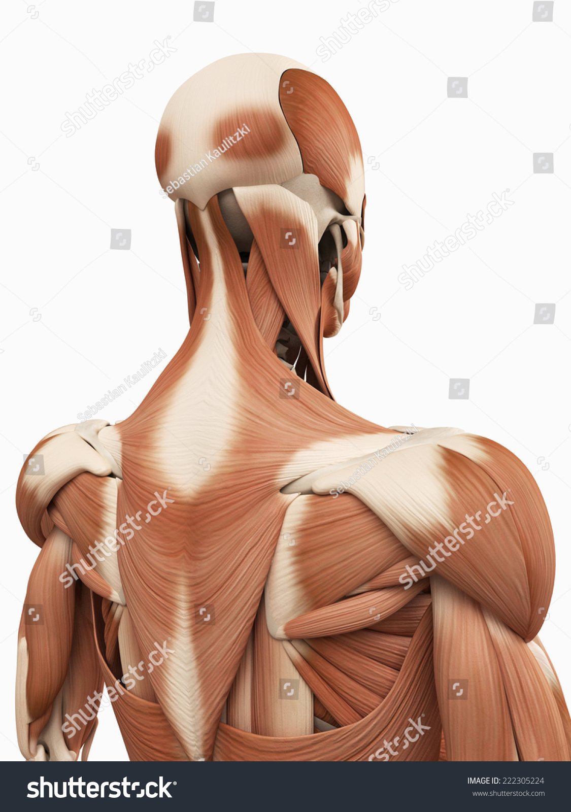
Medical 3d Illustration Upper Back Muscles Stock Illustration 222305224 - Shutterstock from image.shutterstock.com The trapezius and latissimus dorsi muscles connect the upper limb to the vertebral column. The trapezius has upper, middle, and lower groups of fibers. The cervical spine supports the weight and movement of your head and protects the nerves exiting your brain. Anatomy of the upper back. It is like that for several reasons, all of which you can understand by looking at the anatomy of the thoracic spine. Before giving our recommendations for upper back exercises, it's important to first go over the anatomy of the back musculature. There is a set of muscles in the upper back (called the thoracic area) called the spinalis thoracis. The human spine is composed of 4 sections of vertebrae.
The bones of the chest and upper back combine to form the strong, protective rib cage around the vital thoracic organs such as the heart and lungs.
Human musculature bodybuilding infographic muscular system vector human anatomy back muscle anatomy bicep male muscular anatomy human body anatomy female female anatomy muscle hamstrings muscle. Anatomy of the upper back muscles. Oftentimes, patients with upper back pain also have neck pain. It comprises the vertebral column (spine) and two compartments of back muscles; The nervous system of the thorax is a vital part of the nervous system as a whole, as it includes the spinal cord, peripheral nerves, and autonomic ganglia that communicate with and control many vital organs. The trapezius and latissimus dorsi muscles connect the upper limb to the vertebral column. The basic anatomy of your upper back by lindsey mcfadden as you're doing your regular upper back stretching exercises , you're probably wondering about the components of your upper back and why it happens to be the most stable part of your spine. It runs from the neck to the upper back. It is very stiff, and the thoracic spine has a limited range of motion. It extends from the external protuberance of the occipital bone to the lower thoracic vertebrae and laterally to the spine of the scapula. The traps) the latissimus dorsi (a.k.a. The trapezius has upper, middle, and lower groups of fibers. The back is the body region between the neck and the gluteal regions.
This is my video about the muscles of the back. It extends from the external protuberance of the occipital bone to the lower thoracic vertebrae and laterally to the spine of the scapula. The superficial back muscles are situated underneath the skin and superficial fascia. Before giving our recommendations for upper back exercises, it's important to first go over the anatomy of the back musculature. The main superficial muscles of the back are the following:
Diagrams of Back Muscles | 101 Diagrams from www.101diagrams.com The cervical spine supports the weight and movement of your head and protects the nerves exiting your brain. The rib cage also anchors the bones of the head, neck, shoulders, and arms to the trunk of the body. Before giving our recommendations for upper back exercises, it's important to first go over the anatomy of the back musculature. The trapezius and latissimus dorsi muscles connect the upper limb to the vertebral column. The bones of the chest and upper back combine to form the strong, protective rib cage around the vital thoracic organs such as the heart and lungs. Back muscles anatomy here include the trapezius, latissimus dorsi, rhomboid and levator scapulae. Learn to draw the upper back muscles by understanding the anatomical details and forms. They originate from the vertebrae and insert into the scapulae.
This muscle is located on the upper portion of the back anatomy, underneath the trapezius.
Anatomy muscle attachments 12 photos of the anatomy muscle attachments anatomy muscle attachments, anatomy muscle attachments quiz, human anatomy muscle attachments, knee anatomy muscle attachments, shoulder anatomy muscle attachments, human muscles, anatomy muscle attachments, anatomy muscle attachments quiz, human. The trapezius has upper, middle, and lower groups of fibers. It is very stiff, and the thoracic spine has a limited range of motion. The nervous system of the thorax is a vital part of the nervous system as a whole, as it includes the spinal cord, peripheral nerves, and autonomic ganglia that communicate with and control many vital organs. The rib cage also anchors the bones of the head, neck, shoulders, and arms to the trunk of the body. The trapezius and latissimus dorsi muscles connect the upper limb to the vertebral column. They originate from the vertebrae and insert into the scapulae. It comprises the vertebral column (spine) and two compartments of back muscles; The trapezius muscle is a large superficial back muscle that resembles a trapezoid. Both the deltoid and the trapezius are firmly attached to the spine of the scapula. Anatomy of the upper back muscles. The rhomboid muscle is activated as you bring and squeeze your scapula or shoulder blades back and together. See back muscle anatomy stock video clips.
The superficial back muscles are situated underneath the skin and superficial fascia. The rhomboid muscle is activated as you bring and squeeze your scapula or shoulder blades back and together. Anatomy of the upper back. The thoracic spine —also referred to as the upper back or middle back—is designed for stability to anchor the rib cage and protect vital internal organs within the chest. This is my video about the muscles of the back.
Upper Back Workouts from makeoverfitness.com Upper back pain is most commonly caused by muscle irritation or tension, also called myofascial pain. The cervical spine is the top part of the spine. Vertebrae there are 12 vertebrae in the thoracic spine. The lumbar and sacrum region make up the bone of the lower back anatomy. It consists of seven vertebrae. The main superficial muscles of the back are the following: The seventh cervical vertebra, referred to as c7, meets the first of 12 thoracic vertebrae t1 at the base of the neck, a. Both the deltoid and the trapezius are firmly attached to …
The bursa is a small sac of fluid that cushions and.
It is very stiff, and the thoracic spine has a limited range of motion. The basic anatomy of your upper back by lindsey mcfadden as you're doing your regular upper back stretching exercises , you're probably wondering about the components of your upper back and why it happens to be the most stable part of your spine. The human spine is composed of 4 sections of vertebrae. The back is the body region between the neck and the gluteal regions. The lumbar and sacrum region make up the bone of the lower back anatomy. The neck consists of seven cervical vertebrae, the building blocks of the spine. Upper back pain is most commonly caused by muscle irritation or tension, also called myofascial pain. The iliocostalis muscles are furthest from the spine. The rotator cuff is a collection of muscles and tendons that surround the shoulder, giving it support and allowing a wide range of motion. The bursa is a small sac of fluid that cushions and. There is a set of muscles in the upper back (called the thoracic area) called the spinalis thoracis. Anatomy of the upper back. It runs from the neck to the upper back.